Equine Lameness Examination Diagnostics
Equine Diagnostic Nerve & Joint Blocks
Digital Equine Musculoskeletal Ultrasound
Digital Equine Radiography
Digital Equine Musculoskeletal Ultrasound
Digital Equine Radiography
|
A dynamic motion and static physical examination lay the foundation that help determine which diagnostic processes will be of value.
Particularly in cases of limb lameness, nerve or joint "blocks" are used to further localize and rule out a myriad of other possibilities. Blocking can be a vital part of the diagnostic process. You should expect to have this procedure included in your lameness evaluation. After a sterile preparation of the skin called "scrubbing", a small amount of anesthetic, similar to Novocaine that is used by human dentists, is injected under the skin near the nerve or nerve bundles or directly into the joint. The horse is then evaluated in motion to determine if there is improvement due to "blocking" of the pain signal. The amount of time we wait between administration and evaluation depends on which block. We may evaluate the horse right away or we may wait 15 minutes. If there has been no improvement in lameness, we go onto the next level of blocks. The first block will always be the lowest on the limb and we work our way up the limb as needed. Most nerve blocks and some joint blocks do not require bandaging but some joint blocks are protected by a bandage. How Long Does It Take? Blocking can take a half hour or 3 hours, it just depends on how far up the limb we have to go. Occasionally, it is necessary to block again on another day to prove the location of pain has been adequately identified. If multiple blocks have been done, most horses will get intravenous phenylbutasone "IV bute" to minimize tissue inflammation from needle insertion. |
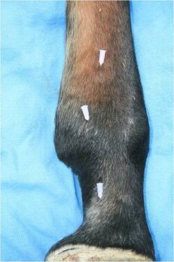
Marking the location of three separate levels of nerve blocks. The lowest one, near the hoof, is usually the starting point unless there is clinical indication that the problem does not involve the structures in the hoof.
|
|
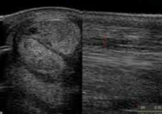
Equine musculoskeletal ultrasongraphy is one of Dr. Morgan's favorite subjects to talk about, to do, to teach, to research. Under the guidance of Dr. Jean-Marie Denoix and the International Society of Locomotor Pathology she has learned how to effectively evaluate every joint, tendon and ligament in the horse from the neck, back and pelvis to the entire front and hind limb using ultrasound.
Morgan Equine has a GE LogicE laptop digital ultrasound machine that is full of features and specialty probes to see detail with extraordinary clarity. We are no longer limited to fuzzy black and white static while ultrasounding the tendons on the back of a cannon bone. Every soft tissue part including the eye, spinal cord and some bone structures can be ultrasounded. Its an extremely sensitive diagnostic tool. |
|
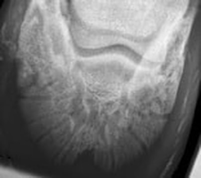
Digital radiographic equipment provides consistent image quality and the ability to find small bone and soft tissue detail on a single image. We also have significantly less exposure of radiation to horses and humans and the time it takes to process information is undeniably one of the most attractive features.
We use an Eklin DR system with a minXray generator. We can image limbs, the skull, neck and portions of the back. Pelvic images require a much bigger machine to capture images in the standing horse. Most horses tolerate radiography well. Depending on the nature of the horse; sedation may or may not be necessary to take diagnostic quality images. Images are obtained by capturing x-rays on a plate that is connected to a computer that processes information with complex logarithms. The best part for clients is that images are taken, viewed and interpreted immediately during the visit. Dr. Morgan will interpret the images and explain any unusual findings and their significance. She will take her time making sure you are confident in your understanding of the findings. It is important to remember that abnormalities found on radiographs might not necessarily be a problem. It can take about a half hour to an hour to radiograph a horse. Evaluation of images and consultation will take as long as you need to understand the issues. Typically, after radiographs are evaluated there are two possibilities. Either more information will be necessary to come to a conclusion or a therapeutic plan begins to develop. Radiography can be diagnostic for many conditions, but remember it's use is typically confined to evaluating bone structures. Many people ask about Pre-purchase radiography. What should I have done? How many? How much does it cost? Taken by Who? Can Dr. Morgan evaluate them? The answers to those questions are different for each situation. Each horse has a specific job it needs to fulfill. It might take 4 radiographs or 36 to make a decision. A general rule of thumb is to set aside 5% of the cost of the horse for a pre-purchase exam and diagnostics including radiography. Images or links to images can be sent to morganequine@gmail.com. |
|